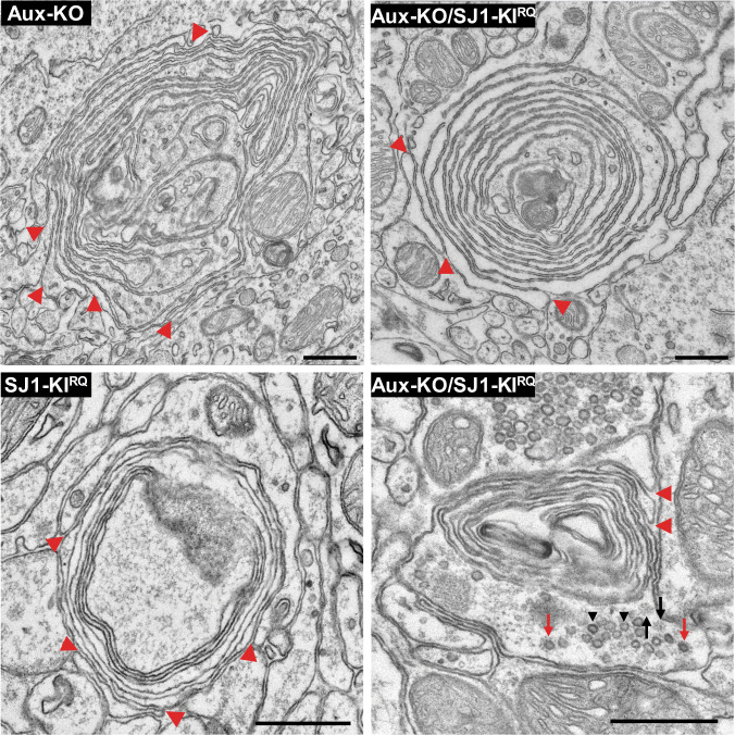
Fig. 5. EM micrographs showing multilayered onion-like membrane structures in the dorsal striatum of Aux-KO, SJ1-KIRQ, and Aux-KO/SJ1-KIRQ mice.
These structures, which are positive for TH immunoreactivity (see Supplementary Fig 6), appear to result from invaginations of the plasma membrane as indicated by red arrowheads. Note in the bottom right field presence of a cluster of SVs (black arrowheads) and scattered CCVs (red arrows) and empty clathrin cages (black arrows), confirming that these structures represent dystrophic changes of nerve terminals. Scale bar: 500 nm.

