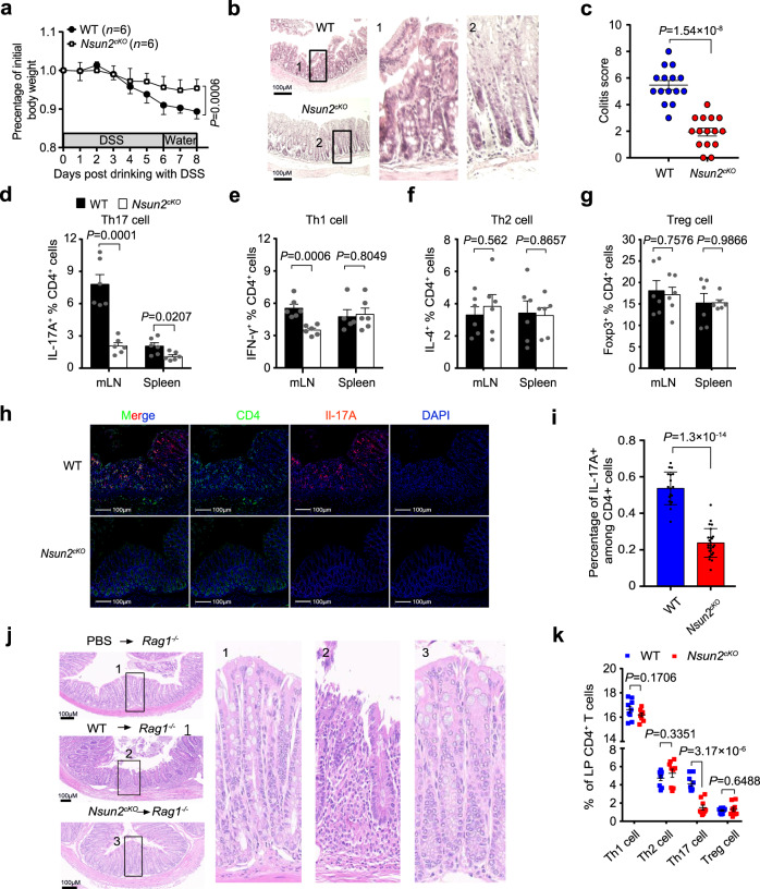
Fig. 4. Targeting Nsun2 in T cells ameliorates Th17-mediated autoimmunity.
a–i The phenotype of wild-type and Nsun2cKO mice under DSS-induced. Littermate wild-type (n = 6) and Nsun2cKO (n = 6) mice were treated with 3% DSS. Body weight changes (a), hematoxylin-eosin (H&E) staining indicated the severity of colons damage and inflammatory infiltration (b), and colitis score (n = 15 image per group) (c). FACS showing the T cell subsets frequencies of mesenteric lymph nodes and spleen, including Th17 cell (n = 6 per group) (d), Th1 cell (n = 6 per group in mLN; n = 5 for WT and n = 6 for Nsun2cKO in spleen) (e), Th2 cell (n = 6 per group) (f), and Treg cell (n = 6 per group) (g). h, i Immunofluorescence assay showing Th17 infiltration in the colons after DSS-induced colitis. Scale bar, 100 μm. Statistical analysis of the frequency of IL-17A+ CD4+ T cells in colonic CD4+ T cells from immunofluorescence image (n = 18 for WT; n = 25 for Nsun2cKO) (i). j, k The phenotype of wild-type and Nsun2cKO naive T cell adoptively transferring into Rag1-/- recipient mice. Representative hematoxylin-eosin staining indicated the severity of colons damage and inflammatory infiltration (j). Flow cytometry analysis of IFNγ+CD4+ T, IL-4+CD4+ T, IL-17A+CD4+ T, and Foxp3+CD4+ T cells frequency in each group recipient wild-type or Nsun2cKOmice (n = 9 per group) (k). All data are representative of at least three independent experiments. P values were analyzed by two-tailed unpaired Student’s t-test (a, c–g, i and k). Error bars represent mean ± s.e.m. Source data are provided as a Source Data file.

