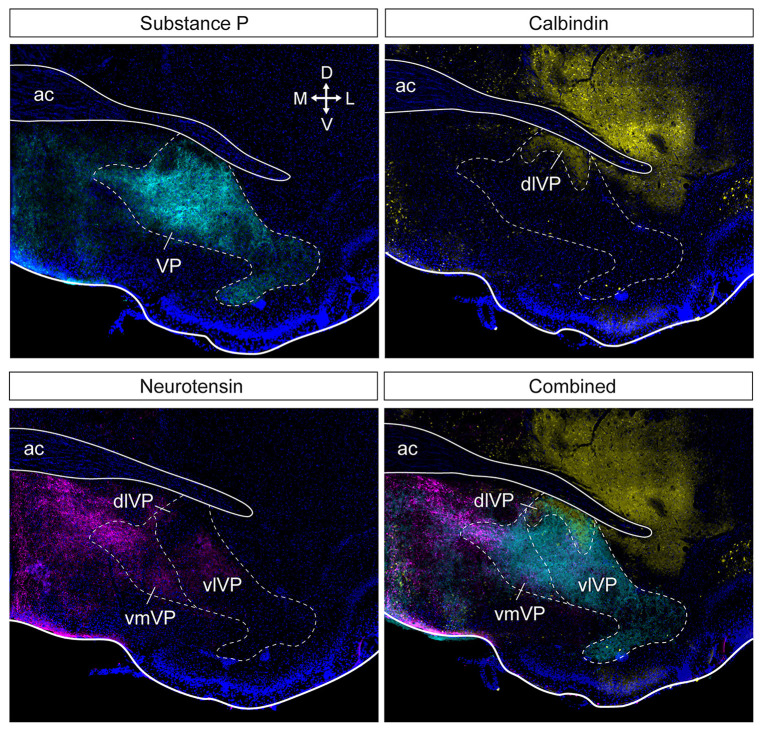
Figure 2.
Subdivision of the ventral pallidum into several functionally and histochemically distinct subregions. The series of adjacent sections (at bregma) were stained for substance P, calbindin, and neurotensin. Substance P (cyan) immunostaining shows the borders of the entire VP (top left). Calbindin (yellow) delineates the dlVP and ventral striatum (top right). Dense neurotensin labeling (magenta) outlines the vmVP and more sparse labeling or lack of labeling indicate the vlVP and dlVP (bottom left). The stains combined show the distinct subterritories of the VP. ac, anterior commissure; dlVP, dorsolateral ventral pallidum; vmVP, ventromedial ventral pallidum; vlVP, ventrolateral ventral pallidum. Arrows indicate the orientation of the brain along the dorsoventral (DV) and mediolateral (ML) axes. Sections are counterstained with DAPI (blue). Figure adopted from Zahm et al. (1996) and Heinsbroek et al. (2020).

