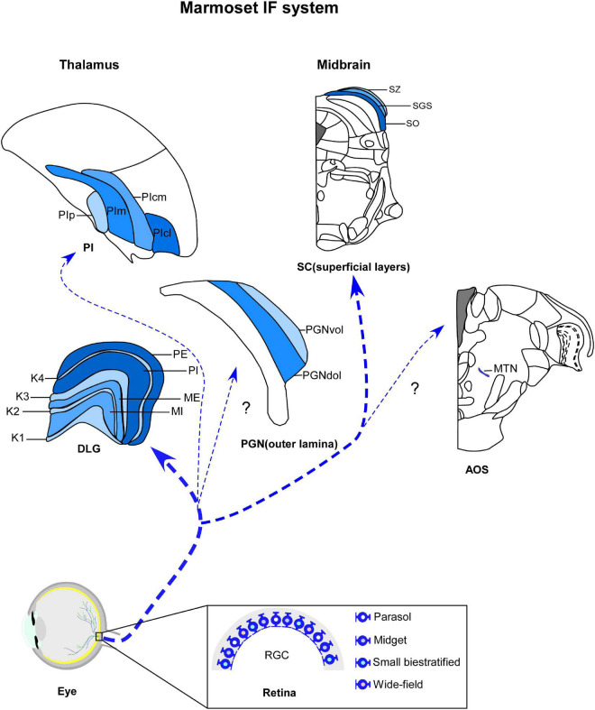
FIGURE 1.
Schematic representation of marmoset image-foming system (blue). Distinct types of retinal ganglion cells (RGCs) bilaterally innervate visual thalamic and midbrain nuclei, such dorsal lateral geniculate nucleus (DLG), inferior pulvinar (PI), outer lamina of the pregeniculate nucleus (PGNol) and superficial layers of superior colliculus (SC). Parasol, midget and small bistratified cells send axonal projections to parvocellular, magnocellular and koniocellular layers of DLG, respectively. SC receives dominant projections from parasol cells. Additional wide-field cells, such as narrow thorny, broad thorny and recursive cell has been described as sending projections to K laminae, CS and PI. The RGC populations that innervates the PGNol, pretectal nuclei and accessory optic nuclei (AOS) still remain unclear. K1–K4, koniocellular layers 1–4; ME, external magnocellular layer; MI, internal magnocellular layer; PE, external parvocellular layer; PGNdol, pregeniculate nucleus dorsal outer lamina; PGNvol, Pregeniculate nucleus ventral outer lamina; PE, internal parvocellular layer; PIcl, central lateral nucleus of the inferior pulvinar; PIcm, central medial nucleus of the inferior pulvinar; PIm, medial nucleus of the inferior pulvinar; Pip, posterior nucleus of the inferior pulvinar; PM, medial pulvinar; PL, lateral pulvinar.

