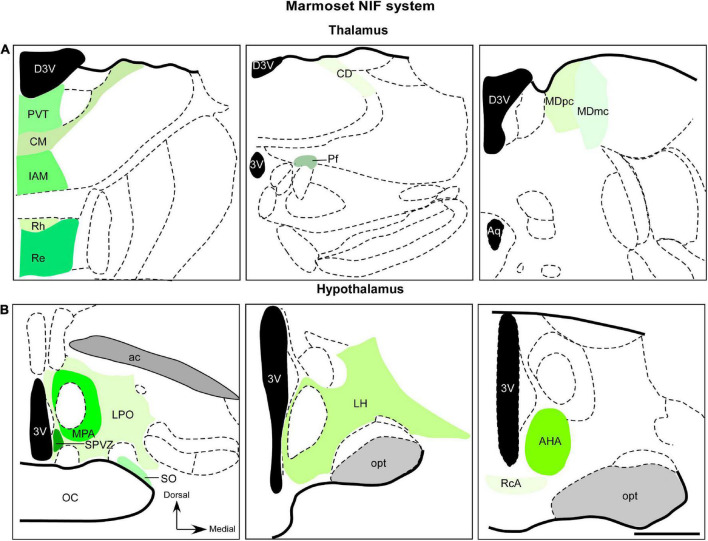
FIGURE 3.
Diagrammatic representation of diencephalic marmoset non-image-foming nuclei (green). Retinal projections were described in mediodorsal nucleus, midline and intralaminar thalamic structures (A) and hypothalamic domains (B). The RGCs subtypes that project to these regions have not yet been elucidated. 3v, third ventricle; ac, anterior commissure; AHA, anterior hypothalamic area; aq, cerebral aqueduct; CD, central dorsal nucleus; CM, central medial nucleus; D3v, dorsal 3v; Iam, inter-antero medial nucleus; LH, lateral hypothalamic area; LPO, lateral preoptic area; MDmc, magnocellular division of mediodorsal nucleus; MDpc, parvocellular division of mediodorsal nucleus; MPA, medial preoptic area; opt, optic tract; Pf, parafascicular nucleus; PvT, paraventricular thalamic nucleus; RcA, retrochiasmatic area; Re, reuniens nucleus; Rh, rhomboid nucleus; SO, supraoptic nucleus; SPVZ; sub paraventricular zone. Scale bar: 500 μm. Adapted from Paxinos et al. (2012).

