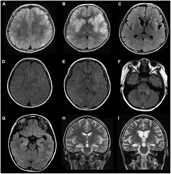
FIGURE 1.
Examples of abnormal brain magnetic resonance imaging (MRI) findings in three patients with pediatric encephalitis/encephalopathy with anti-GAD antibody. Patient 1: A 10-year-old boy with clinical diagnosis of encephalitis with preceding influenza B infection 6 days ago. (A,B) Axial fluid-attenuated inversion recovery (FLAIR) sequences show multiple increased signals in the basal ganglia and cortical/subcortical regions of bilateral cerebral hemispheres (frontal, parietal, temporal lobes). (C) MRI follow-up at 9 months show progressive atrophy with stationary hyperintensities in bilateral putamen, bilateral insular regions, and left temporal tip. Patient 2: A 9-year-old girl with clinical diagnosis of rhombencephalitis with preceding HSV-1 infection 6 days ago. (D–F) Axial FLAIR sequences demonstrate multiple increased signals in the corpus callosum, bilateral insular regions, bilateral posterior capsules, and multiple cortical areas and white matter of bilateral hemispheres (all lobes), the brainstem, and left middle cerebellar peduncles. Patient 3: A 6-year-old girl with clinical diagnosis of febrile infection-related epilepsy syndrome (FIRES) without detected pathogen. (G) An axial FLAIR sequence and (H) a coronal T2 sequence show increased signals in right hippocampus and mesial temporal lobe. (I) Progressive cortical atrophy and right hippocampal atrophy were noted at the 30-month follow-up. GAD, glutamic acid decarboxylase.

