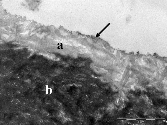
Fig. 3.
In vitro erosion of dentin with 1% citric acid (trial 2). The demineralized dentin (a) had a lower electron density than the unaffected dentin (b). Note the exposed collagen fibrils with a striped pattern and the electron-dense particles (arrow) at the surface. Original magnification: 30,000-fold.

