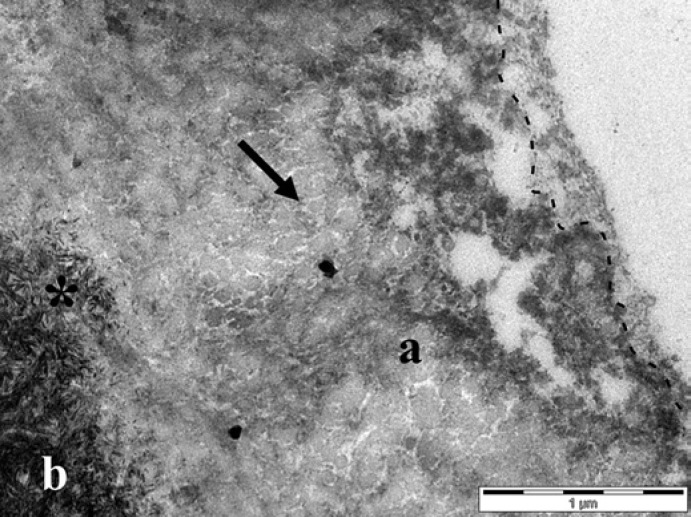
Fig. 5.
Dentin was exposed to the oral cavity for 30 min, eroded with 1% citric acid in vivo, and worn for a further 60 min (trial 3). The demineralized dentin (a) had a lower electron density than the unaffected dentin (b) and was covered by a granular and globular structured pellicle layer. The interface between the pellicle and dentin is marked by a dashed line. Note the granular material infiltrating the hollow spaces of the demineralized dentin (arrow) and the disclosed hydroxyapatite crystals at the intermediate zone (*). Original magnification: 30,000-fold.

