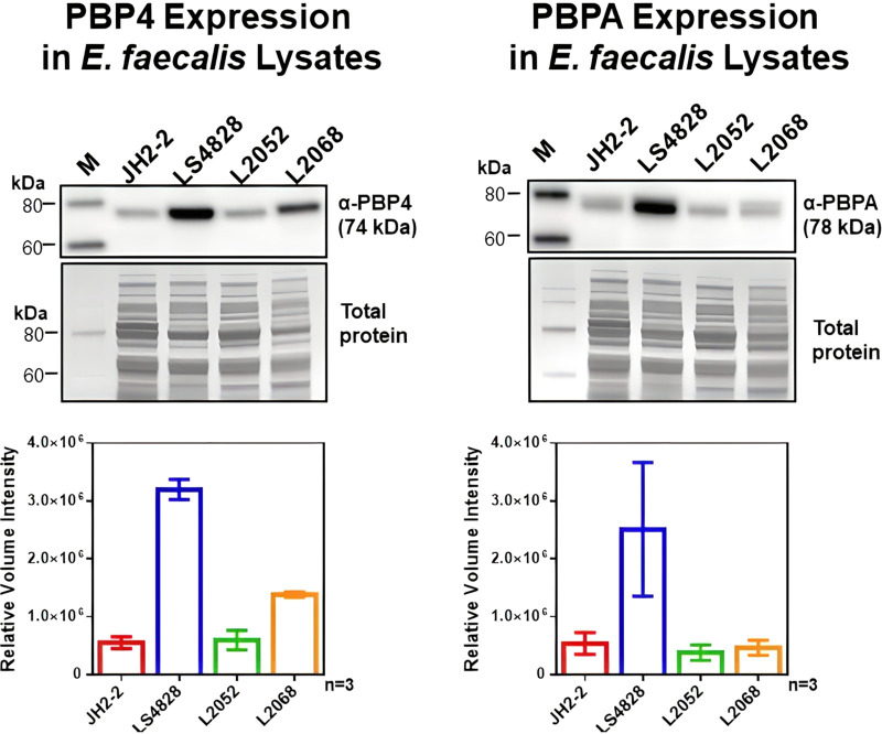
FIG 2.
Quantitation of PBP4 and PBPA expression by Western blot analysis. E. faecalis cells grown in BHI broth to exponential phase were processed for SDS-PAGE and transferred to PVDF membranes for immunoblot detection of PBP4 or PBPA using a custom polyclonal antibody. The graphs below each blot represent expression levels from 3 biological replicates. These data were from normalized densitometry analysis after Coomassie blue R-250 staining of total proteins in the blots. M, molecular weight marker.

