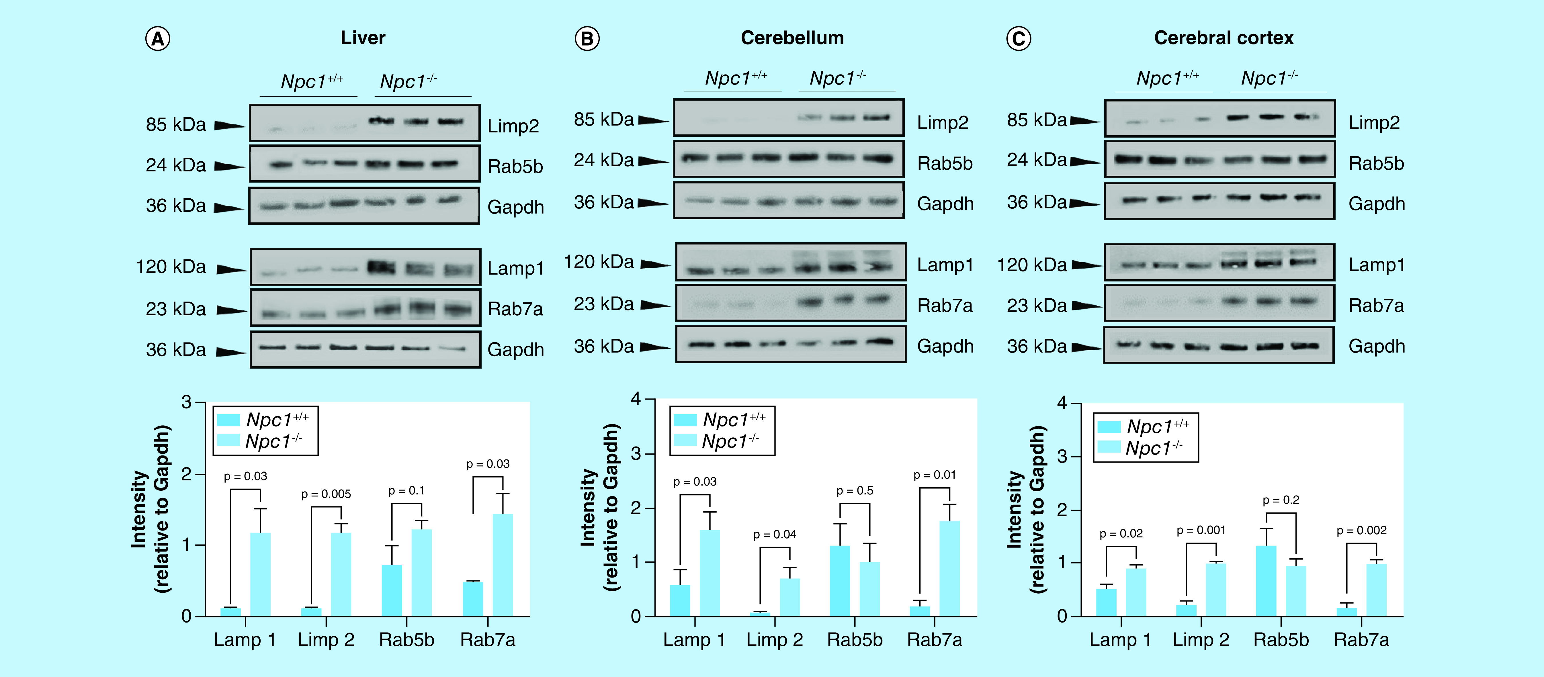
Figure 4. . Western blot analysis of lysosome and phagosome/endosome markers in liver, cerebellar, and cerebral cortex tissue from 11-week mice from the symptomatic NPC1 mouse model.

Individual lysates (N = 3 for each genotype) from 11-weelk Npc1+/+ and Npc1-/- animals from the (A) liver, (B) cerebella, and cerebral cortex (C) were subject to electrophoresis and western blotting of ras-related binding protein 5b (Rab5b), ras-related binding protein 7a (Rab7a), lysosome membrane protein 2 (Limp2) and lysosome-associated membrane glycoprotein 1 (Lamp1). Intensities are reported as relative to Gapdh. Significance was determined using an unpaired t-test with Welch’s correction.
