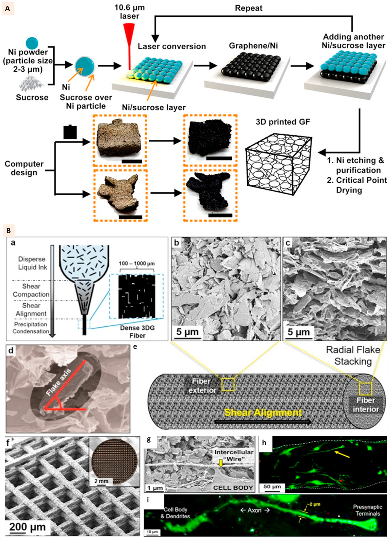
Fig. 2.

(A) Schematics showing the 3D printing of graphene foams and images of the 3D printed graphene foams before and after dissolving the nickel layer. Scale bar: 5 mm. (Adapted with permission from Ref. [30], Copyright 2017, American Chemical Society). (B) ‘a’ shows the liquid nature of graphene-PLG ink prior to application of pressure and flow, ‘b’, ‘c’, ‘d’, and ‘e’ show the images of graphene flake alignment along the exterior of extruded fibers and flake stacking within the fibers. ‘f’ shows the SEM and optical (inset) image of 3D printed structure. ‘g’ shows the SEM image of hMSC connecting via a small segment of long intercellular wire on the scaffolds at day 7. ‘h’ and ‘i’ show the confocal images of cells with neuronal structures growing on the scaffolds (Adapted with permission from Ref. [31], Copyright 2015, American Chemical Society).
