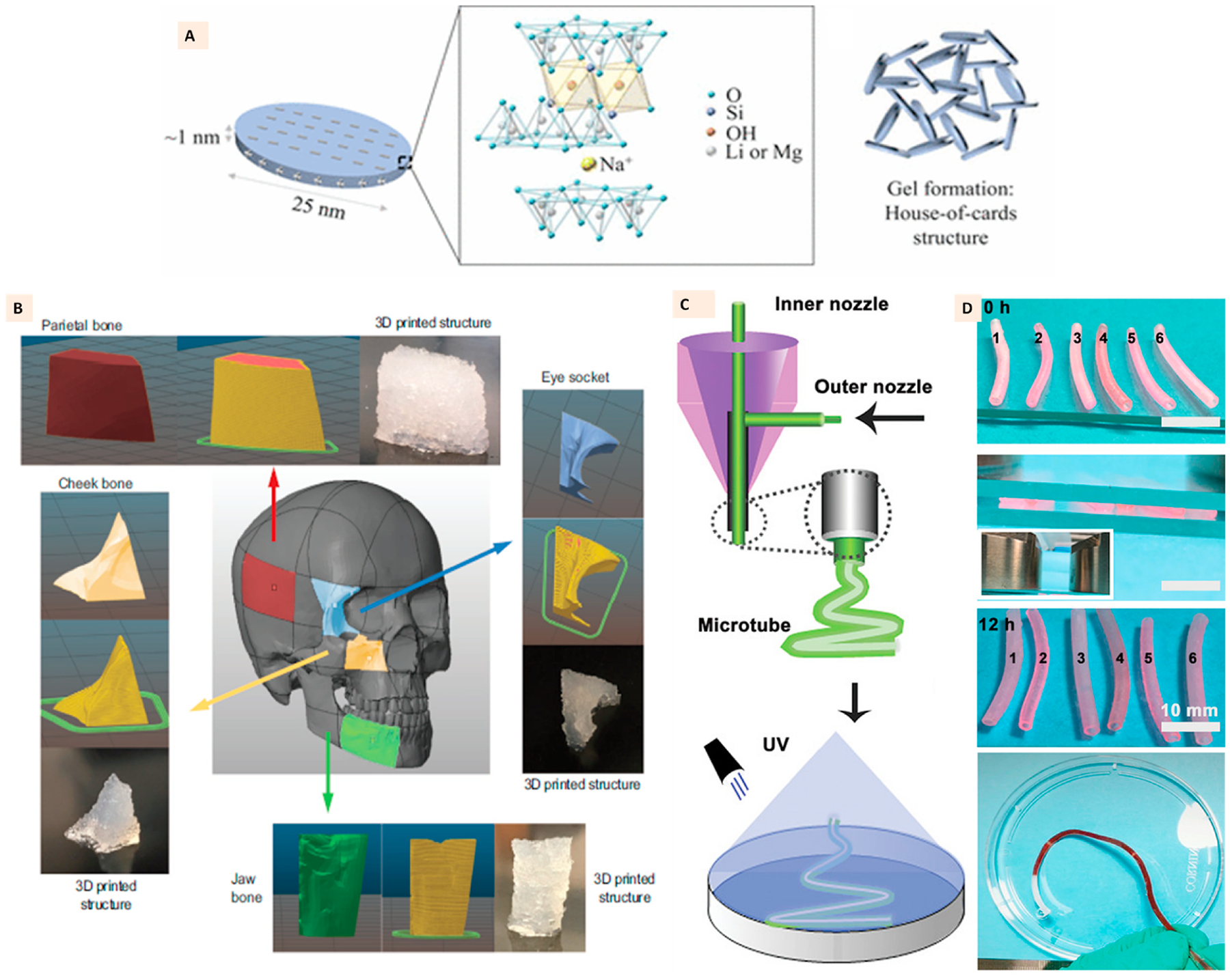
Fig. 6.

(A) Single layer of Laponite clay with its structural formula and house-of-cards arrangement of several layers in laponite gel. (Adapted with permission from Ref. [96], Copyright 2017, American Chemical Society). (B) 3D printed anatomical structures with Laponite-GelMa-kappa carrageenan ink demonstrating high printing fidelity and high design precision to reproduce the anatomical craniomaxillofacial defects. (Adapted with permission from Ref. [100], Copyright 2020, John Wiley and Sons). (C) Coaxial extrusion printing of Laponite, GelMa, and NAGA ink to develop hydrogel microtubes with high strength, perfusability, and endothelialization, (D) Photographs of printed microtubes with the demonstration of perfusion through tubes. (Adapted with permission from Ref. [103], Copyright 2020, John Wiley and Sons).
