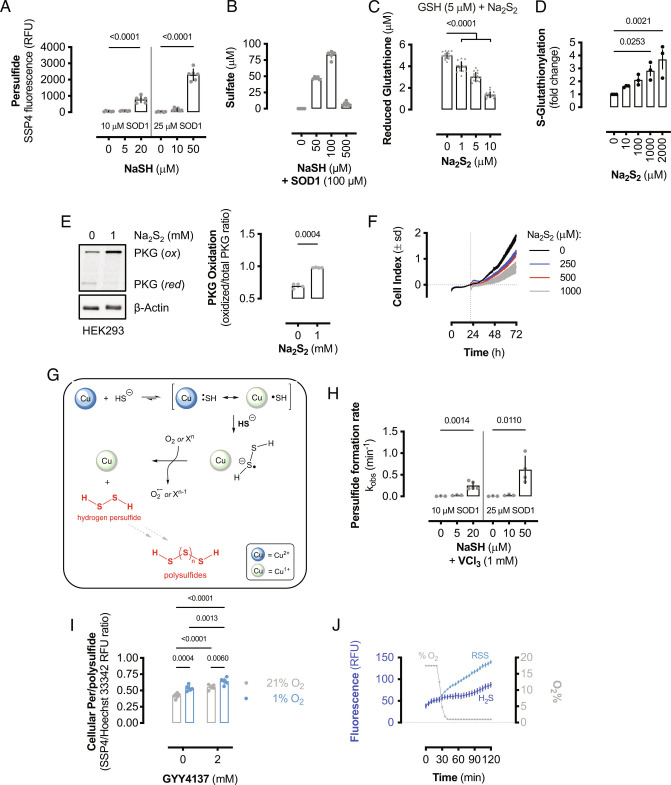
Fig. 3.
SOD1 oxidizes excess H2S to RSS. (A) RSS formation from the oxidation of NaSH by human SOD1. Bars represent mean values (±SEM; n = 6 reactions). Significance calculated by one-way ANOVA with Dunnett’s test. (B) Sulfate formation from the oxidation of NaSH by 100 μM human SOD1. Bars represent mean values (±SEM; n = 5 to 6 reactions). (C) Reduced glutathione remaining after reacting GSH (5 μM) with Na2S2 at the indicated concentrations. Bars represent mean values (±SEM; n = 18 wells). Significance calculated by one-way ANOVA with Dunnett’s test. (D) Quantification of protein S-glutathionylation from MCF10A cells treated with Na2S2. Bars represent mean values (±SEM; n = 3 wells) and significance calculated by one-way ANOVA with Dunnett’s test. (E) Immunoblot of relative reduced and oxidized PKG and β-actin expression in HEK293 cells treated with or without 1 mM Na2S2. Bars represent mean oxidized PKG fraction (±SEM; n = 4 wells). (F) Cellular proliferation as measured by electrical impedance (cell index). HEK293 cells were cultured for 24 h before addition of Na2S2 stock solutions and cellular proliferation monitored for 48 h. Data shown are mean cell index values (±SD; n = 3 wells). (G) Proposed chemical mechanism for SOD1-catalyzed RSS formation from excess sulfide. The reaction proceeds via a disulfide radical anion intermediate. (H) Observed rate constants for the formation of persulfide from the anaerobic reaction of limiting or excess sulfide with SOD1 in the presence of 1 mM VCl3. Bars represent mean rate constants (±SEM; n = 3 to 5 reactions). Significance calculated by one-way ANOVA with Dunnett’s test. (I) Relative cellular persulfide and/or polysulfide formation in HEK293 cells cultured with GYY4137 or vehicle in normoxic (21% O2) or anaerobic conditions (1% O2). Bars represent mean values (±SEM; n = 6 wells). Significance calculated by two-way ANOVA with Sidak’s test. (J) Atmospheric O2 content and relative fluorescence of wild-type yeast loaded with either a H2S- or RSS-specific probe cultured at 30 °C. Data shown are mean values (±SEM; n = 9 to 10 wells).

