Fig. 5.
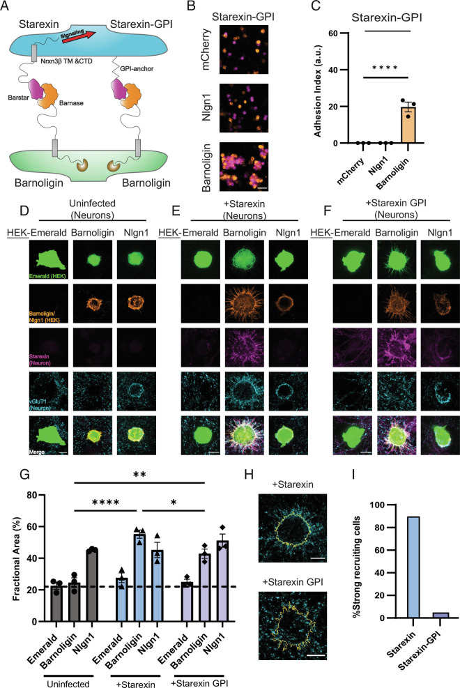
Intracellular signaling is required for Starexin to direct presynaptic organization. (A) Cartoon depicting the design of the Starexin-GPI construct. (B) HEK293F cells expressing Starexin GPI (purple, all panels) specifically form an adhesion complex only with cells that are expressing Barnoligin (orange, Bottom) and not mCherry alone or Nlgn1 (both orange, Top and Middle, respectively). (C) Quantification of B. (D) As before, Barnoligin-expressing HEK cells do not induce presynaptic accumulation in uninfected neurons. (E) Starexin expressed in neurons directs synapse organization to the surface of Barnoligin-expressing co-cultured HEK cells. (F) Neuronally-expressed Starexin GPI accumulates on the surface of Barnoligin-expressing HEK cells as Starexin does, but the characteristic accumulation of vGluT1 is absent. (G) Quantification of D–F (gray, blue, lavender, respectively). Although characteristic halos of vGluT1 are absent from Starexin GPI-expressing neurons, careful quantification reveals a significant increase in presynaptic specializations co-incident with Barnoligin-expressing HEK cells. (H) Close-up detail of two Barnoligin-expressing HEK cells co-cultured either with Starexin (Top) or Starexin-GPI (Bottom). Cell outlines are shown in yellow. (I) Quantified fraction of Barnoligin-expressing cells co-cultured with Starexin or Starexin GPI-expressing neurons demonstrating characteristic recruitment halos. Statistical comparisons in C and G made with two-way ANOVA with Dunnet’s multiple comparison correction (*P < 0.05; ***P < 0.001; ****P < 0.0001).

