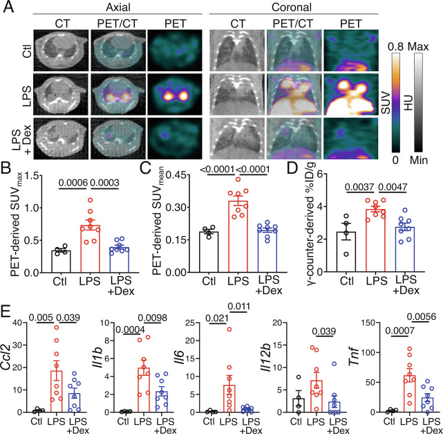
Fig. 3.
Effect of dexamethasone treatment on the lung uptake of [64Cu]NODAGA-CG34 in LPS-induced experimental lung injury. (A) Representative axial (Left) and coronal (Right) CT, PET, and co-registered PET/CT on day 2 post-PBS or LPS treatment. Images were acquired ~90 min after intravenous [64Cu]NODAGA-CG34 injection in control (Top row) and LPS-treated mice after receiving (Bottom row) or not receiving (Middle row) dexamethasone. Corticosteroid treatment reduced radiotracer uptake by PET, although areas of airspace opacities were still frequently observed on CT. (B and C) In vivo PET-derived quantification of tracer uptake demonstrates ~46% decrease in lung SUVmax and ~41% decrease in SUVmean in LPS-injured mice receiving dexamethasone compared to mice treated with LPS only (N = four males and four females per group). The lung radiotracer uptake in dexamethasone-treated mice approached that of control mice (N = two males and two females for control mice). (D) Quantification of lung radiotracer uptake by γ-counting confirms a similar pattern with 28% decreased [64Cu]NODAGA-CG34 uptake in LPS-injured mice treated with dexamethasone, when compared to mice receiving only LPS. (E) The mRNA expression of select inflammatory markers is significantly reduced in dexamethasone-treated, compared to untreated, LPS-injured lungs. mRNA transcript levels are normalized to the geometric mean of Rn18s, the housekeeping gene. SUVmax and SUVmean, and %ID/g values represent the average values of the left and right lungs for each mouse. PBS = phosphate-buffered saline; LPS = lipopolysaccharide; Dex = dexamethasone. Data are expressed as the mean ± SEM. Statistical significance between groups was calculated using a one-sided ANOVA with a post hoc two-tailed Fisher’s exact test.

