Fig. 4.
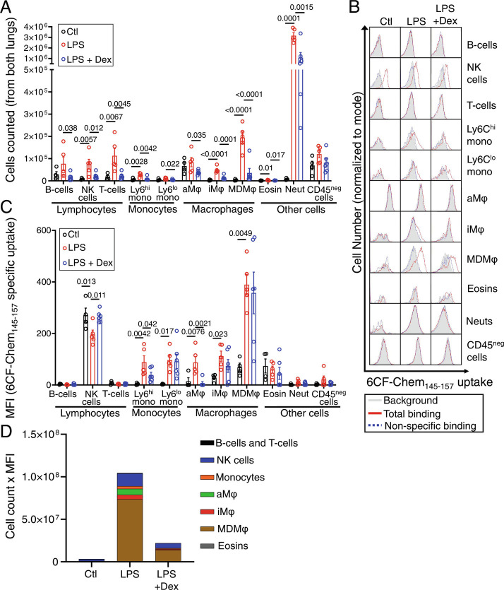
Flow cytometry identification of CMKLR1 expressing cells in healthy and LPS-injured lungs. (A) The absolute number of most immune cells within the lungs is significantly increased at day 2 post-intratracheal instillation of LPS. (B) Representative histograms showing specific uptake of a CMKLR1-targeted fluorescent ligand, 6CF-Chem145–157 (100 nM), by various immune cells in the lungs of mice treated with PBS, LPS, or LPS plus dexamethasone (gray: background/autofluorescence; red: total-binding of 6CF-Chem145–157 in the absence of Chem145–157; blue: non-specific binding of 6CF-Chem145–157 in the presence of 10 µM Chem145–157). (C) Specific 6CF-Chem145–157 uptake (total minus non-specific/blocked) in different cell subsets was quantified by mean fluorescent intensity (MFI), as a proxy for the uptake of CMKLR1-targeted tracer ([64Cu]NODAGA-CG34), in LPS-injured vs. control mice. Monocyte-derived macrophages, interstitial macrophages, and monocytes (Ly6Chi and Ly6Clo) demonstrated the largest increases in 6CF-Chem145–157 uptake in LPS-induced experimental lung injury. NK cells had significant 6CF-Chem145–157 uptake during steady state, but the uptake was not further induced by lung injury. (D) A stacked-bar graph summary of the cell count multiplied by the cellular uptake (MFI) of 6CF-Chem145–157 highlights that monocyte-derived macrophages are the major contributors to the tracer uptake (~70%) due to a marked increase in their abundance and significant induction of 6CF-Chem145–157 uptake per cell in mice with LPS-induced ALI compared to control or dexamethasone-treated mice. CD45-negative cells and neutrophils are omitted from this graph due to their negligible specific 6CF-Chem145–157 uptake. N for PBS group = two male and two female mice; N for LPS group: three male and two female mice; N for LPS + dexamethasone group: three male and three female mice. aMφ: alveolar macrophages; Eosins: eosinophils; iMφ: interstitial macrophages; MDMφ: monocyte-derived macrophages; NK cells: natural killer cells; Neut: neutrophils. Data are expressed as the mean ± SEM. Statistical significance between groups was calculated using a one-sided ANOVA with a post hoc two-tailed Fisher’s exact test.

