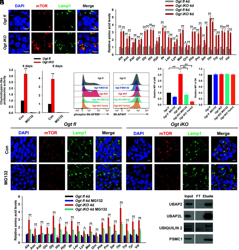
Fig. 6.
OGT deficiency promotes the translocation of mTOR by increasing proteasome activity. (A) Immunohistochemistry of Ogt fl and Ogt iKO mESCs treated without or with 4-OHT, respectively, for 6 d. Cells were stained with antibodies against mTOR and Lamp1. Nucleus staining: DAPI (blue). Scale bar: 10 μm. (B) Relative amino acid levels in Ogt fl and Ogt iKO mESCs treated without or with 4-OHT, respectively, for 4 or 6 d. Data are shown as mean ± SD (N = 3). (C) Chymotrypsin-like proteasome activity in Ogt fl and Ogt iKO mESCs treated without or with 4-OHT, respectively, for 4 or 6 d. Data are shown as mean ± SD (N = 3). (D) Flow cytometry analysis of phospho-S6 (Left) and total S6 (Right) ribosomal protein in Ogt fl and Ogt iKO mESCs treated without or with 4-OHT, respectively, for 6 d and then with or without 10 μM MG132 or 200 nM Torin2 for 2.5 h. (E) Relative mean fluorescence intensity (MFI) of phospho-S6 and S6 ribosomal protein in three independent experiments similar to that shown in (D). (F) Immunohistochemistry of Ogt fl and Ogt iKO mESCs treated without or with 4-OHT, respectively, for 6 d and then with or without 10 μM MG132 for 2.5 h. Cells were stained with antibodies against mTOR and Lamp1. Nucleus staining: DAPI (blue). Scale bar: 10 μm. (G) Relative amino acid levels in Ogt fl and Ogt iKO mESCs treated without or with 4-OHT, respectively, for 4 d and then with or without 10 μM MG132 for 2.5 h. (H) Western blot of UBAP2, UBAP2L, ubiquilin 2, and PSMC1 proteins after WGA pull down of cell lysates from Ogt fl mESCs. FT: Flow Through.

