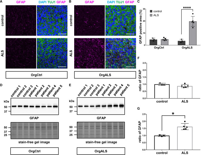
Fig 4. ALS patient-derived protein extract injection induces astrogliosis in cerebral organoids.
(A and B) Immunofluorescence images of GFAP and TUJ1 staining OrgCtrl (A) or OrgALS cerebral organoids (B) at 8 weeks p.i. of protein extract from control (control 1) or ALS (patient 3). DAPI was used for counterstaining of nuclei. Scale bars = 50 μm. (C) Quantification analysis of immunofluorescence images of GFAP staining OrgCtrl and OrgALS cerebral organoids at 8 weeks p.i. of protein extract from individual controls (control 1–3) or ALS cases (patient 1–5) represented in (A) and (B). Percentages of GFAP-positive area were measured with ImageJ software. Bar plots show mean ± SD with individual points representing a different proteins extract. Differences were evaluated by two-way ANOVA with Tukey’s multiple comparisons test. ****p<0.001. (D and E) Western blot analysis of GFAP expression in OrgCtrl (D) or OrgALS cerebral organoids (E) at 8 weeks p.i. of protein extracts from individual controls (control 1–3) or ALS cases (patient 1–5). A stain-free gel image was used for protein loading controls. (F and G) Densitometric quantification of Western blot analysis in (D) and (E), respectively, using ImageJ software. Each data point was obtained by normalization to bands in stain-free gel blot images. Bar plots show mean ± SD with individual points representing a different protein extract from controls (control 1–3) and ALS (patient 1–5). Differences were evaluated by unpaired t-test. *p<0.05.

