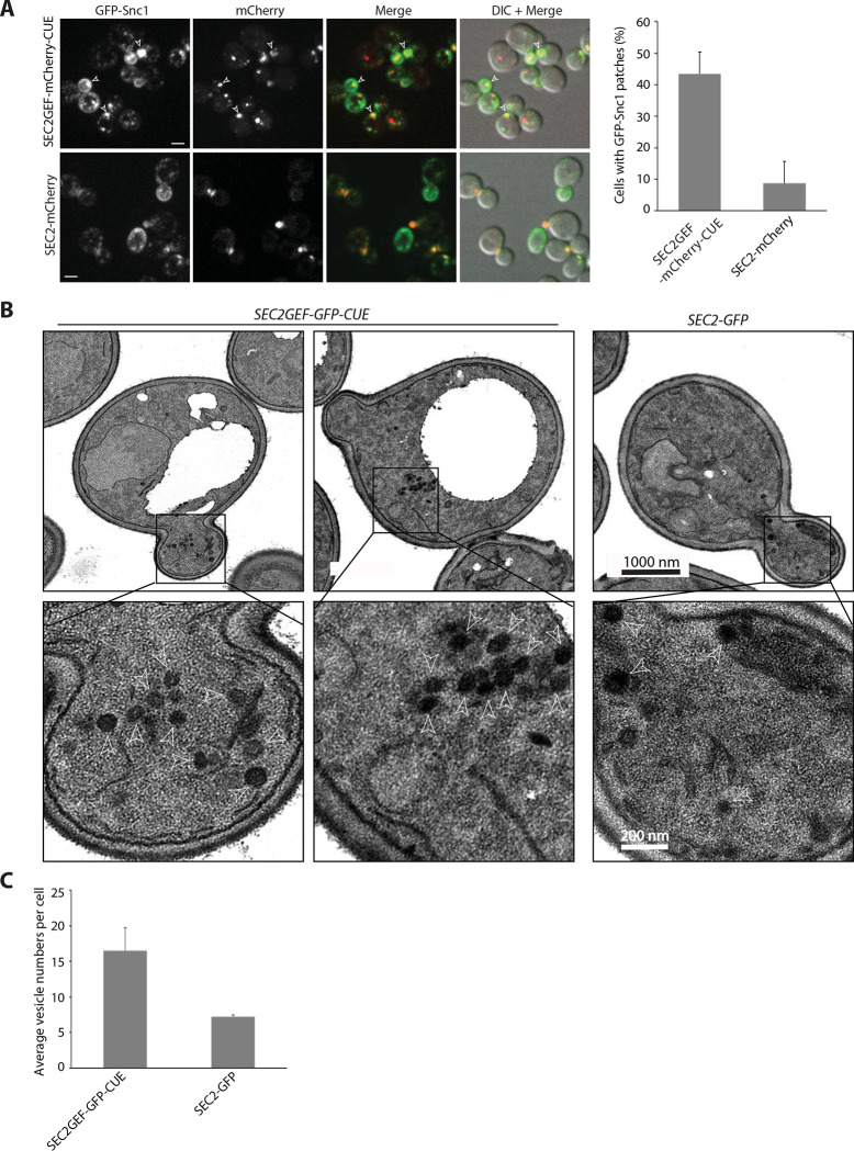
Figure 5.
The SEC2GEF-CUE mutant has mild secretion defects shown by GFP-Snc1 localization and EM (A). Left panel shows representative fluorescence images of GFP-Snc1, the mCherry channel, a merge of the two channels and overlay DIC with merged images in SEC2-mCherry or SEC2GEF-mCherry-CUE cells grown to early log phase in SC medium at 25°C. Open arrowheads point to accumulated GFP-Snc1 patches. Bars, 5μm. The percentage of cells that contained GFP-Snc1 patches in indicated strains was quantified (right panel). The error bars represent the SD from three independent experiments. (B). Thin-section EM images from the strains indicated were prepared as described in Materials and Methods. The cells were fixed in potassium permanganate. Representative images at 10000x magnification are shown on top panels. The Expanded boxed regions from top panels are shown on bottom panels. Arrowheads indicate vesicles and Scale bars are shown as indicated. (C). Quantitation of secretory vesicles per cell are shown. Over 100 cells were analyzed for each strain.

