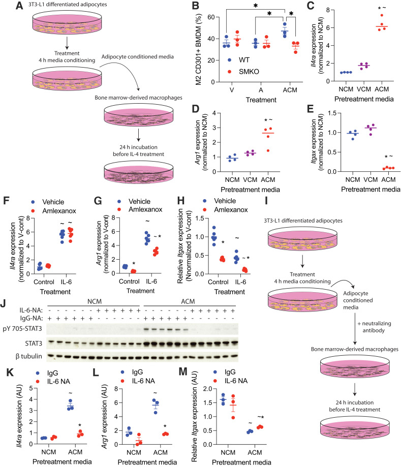
Figure 3.
Adipocyte-secreted IL-6 sensitizes macrophages to IL-4. Adipocyte conditioned media was generated by treating adipocytes with 100 μmol/L amlexanox in RPMI medium for 4 h. Direct treatment of BMDMs with amlexanox was also performed with 100 μmol/L amlexanox. A: Schematic of adipocyte media conditioning and treatment of BMDMs. B: Percent CD301+ staining of F4/80, CD11b dual-positive BMDM treated with amlexanox directly, or ACM (n = 3 per group). *P < 0.05, comparison indicated by line. C–E: Gene expression in BMDMs pretreated with NCM, VCM, or ACM for 24 h before the addition of IL-4 for another 24 h (n = 4 per group). *P < 0.05 ACM vs. VCM; ∼P < 0.05 ACM vs. NCM. F–H: Gene expression in BMDMs treated with 50 ng/mL IL-6 with and without amlexanox, normalized to the VCM (n = 6 per group). *P < 0.05 vehicle vs. amlexanox; ∼P < 0.05 control vs. IL-6. I: Schematic of adipocyte media conditioning with neutralizing antibodies and administration to BMDMs. J–M: BMDMs treated with VCM or ACM in which IL-6 was neutralized with IL-6NA or IgG control. *P < 0.05 IgG vs. IL-6NA; ∼P < 0.05 ACM vs. NCM. J: Western blot analysis of pY705 STAT3; β-tubulin serves as a loading control (n = 3 per group). Statistical significance determined by post hoc analysis after significant ANOVA. A, amlexanox; AU, arbitrary unit; V, vehicle; V-cont, vehicle control.

