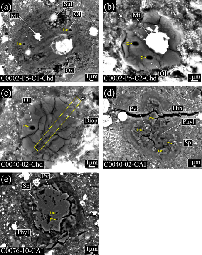
Fig. 1. Backscattered electron (BSE) images of three chondrule-like objects and two CAIs in the Ryugu samples analyzed for oxygen isotopes.

a C0002-P5-C1-Chd, b C0002-P5-C2-Chd, c C0040-02-Chd, d C0040-02-CAI, and e C0076-10-CAI. SIMS analysis spots are shown by the vertex of an open triangle. The rectangle area drown by the dashed line in panel (c) corresponds to the region extracted by the FIB sectioning. Ol, olivine; Mt, Fe-Ni metal; Sul, Fe-sulfide; Ox, oxide; Diop, diopside; Sp, spinel; Hib, hibonite; Pv, perovskite; Phyl, phyllosilicates.
