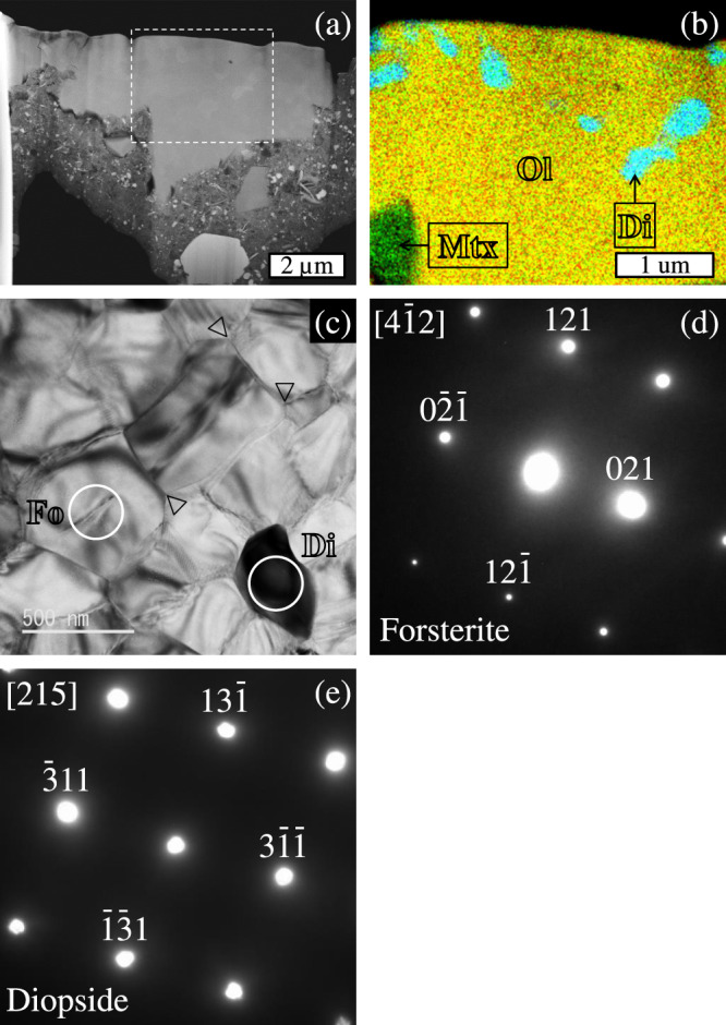
Fig. 2. Sub-micron structures of C0040-02-Chd.

a A high-angle annular dark-field (HAADF)-STEM image of the FIB section from C0040-02-Chd, b a combined elemental map in Mg (red), Si (green), and Ca (blue) X-rays of a rectangle area drawn with the dashed line in panel (a), c a bright field (BF)-TEM image of olivine and diopside in an area of C0040-02-Chd, and d, e selected-area electron diffraction (SAED) patterns from forsterite along the [42] zone axis and diopside along the [215] zone axis. Abbreviations in panel (b): Ol, olivine; Di, diopside; Mtx, matrix. The 120° triple junctions are indicated by the vertex of an open triangle in panel (c). Circled areas in panel (c) represent analysis spots of electron diffraction on forsterite (Fo) and diopside (Di).
