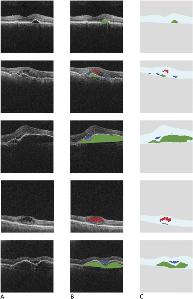
Fig. 1.

A. Scans from the home OCT device. B. Ground-truth labeling: scans from the home OCT device showing manual human grading/delineation of IRF (red), SRF (blue), and sub-RPE fluid (green). C. Scans from the home OCT device processed using the automated analysis software showing retinal layer and fluid segmentation—IRF (red), SRF (blue), and sub-RPE fluid (green).
