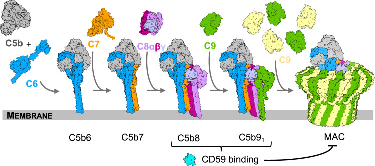
Fig. 1. Schematic of MAC assembly.
The MAC pore is formed from the sequential and stepwise assembly of complement proteins: C5b, grey; C6, blue; C7, orange; C8α, pink; C8β, magenta; C8γ, light purple; C9, alternating monomers are yellow and green. During assembly, complement proteins undergo dramatic structural re-arrangements in which two helical bundles within their MACPF domains unfurl into membrane-inserting β-hairpins. CD59 (cyan) binds at two stages of this assembly process (C5b8 and C5b9) to block membrane perforation and C9 polymerization. Images are rendered from structural models. C5b6 and all MAC assemblies: PDB ID: 6H03; Soluble forms of complement proteins are derived from C6: PDB ID: 3T5O; C8: PDB ID: 3OJY; C9: PDB ID: 6CXO. C7 was derived from an AlphaFold2 prediction: AlphaFold Protein Structure Database P10643.

