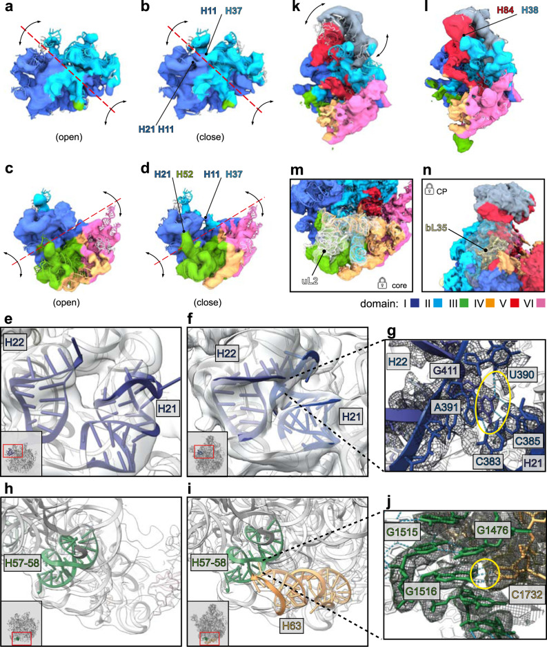
Fig. 5. Tertiary contacts between rRNA helices.
3D variability maps of states d12 (a, b), d136 (c, d), d126-CP (k, l). First (left) and last (right) reconstructed frame of each state is shown. Maps were filtered to 8 Å and color-coded according to the six architectural domains of 23S rRNA. Red dotted lines and black arrows indicate axis and directions of observed global movements, respectively. Stabilizing contacts in closed conformations are indicated by black arrows. Interacting regions are labeled. Cryo-EM maps (transparent gray) and PDB models corresponding to rRNA helices H21 and H22 (blue), H57-58 (green), or H63 (orange), respectively. Local snapshots of state C− (e, h) and state C-CP_L2/L28 (f, i) contain thumbnails with red rectangles indicating the region of interest. g, j close-ups of regions of interest in state C-CP_L2/L28. Yellow circles highlight H-bond interactions (light blue dotted lines) between C385-G411 (g) and G1516-C1732 (j). k, l State d126-CP exhibiting flexibility in the CP occupying a more upright (k) or more inclined position towards the interface (l). Binding sites of L-proteins uL2 (state C_L2) (m) and bL35 (state C-CP-H68) (n).

