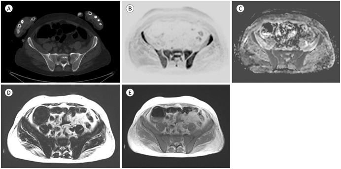
Fig. 3. A 52-year-old female patient with a diffuse pattern of multiple myeloma.
A. CT shows no definite bone lesions in the pelvis and sacrum.
B. Whole-body MR inverted diffusion-weighted image (b-value = 900 sec/mm2) shows diffuse but increased bone marrow signal change compared with that of the soft tissue.
C. The corresponding apparent diffusion coefficient map shows an increased apparent diffusion coefficient value (approximately 700–800 µm2/sec) compared to the normal bone marrow (less than 600–700 µm2/sec).
D, E. T1-weighted Dixon fat-only (D) and in-phase (E) images show decreased signal intensity at the sacrum and pelvic bone, suggesting a diffuse pattern of multiple myeloma involvement.

