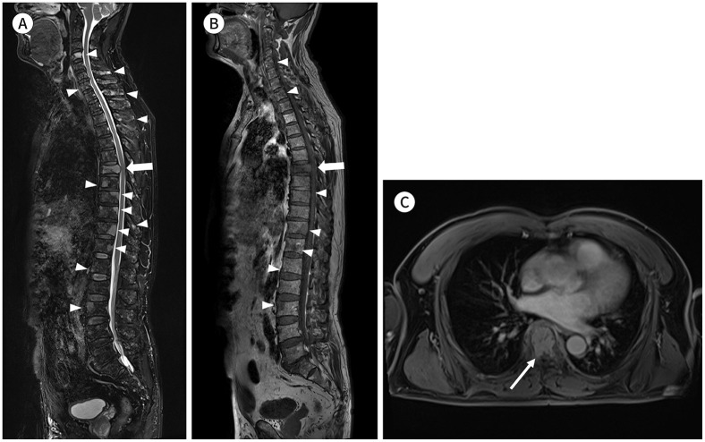
Fig. 4. A 61-year-old male patient with multiple myeloma.
A-C. Sagittal fat-suppressed T2-weighted (A) and T1-weighted (B) images and axial contrast-enhanced fat-suppressed T1-weighted image (C) show the decreased height of the T7 vertebral body with diffuse bone marrow signal change and anterior epidural mass formation abutting the spinal cord (arrows), suggesting a pathologic compression fracture. Other multifocal bone lesions are visible in the whole spine (arrowheads).

