Figure 1.
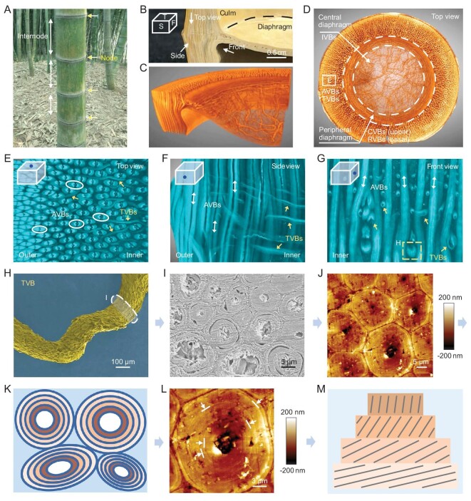
Morphology and hierarchical fiber arrangement of the bamboo node. (A) Digital image of moso bamboos (Phyllostachys edulis). Yellow arrows and white arrows show nodes and internodes of one bamboo, respectively. (B) Partial node showing pre-defined view directions (abbreviations: T, top view; S, side view; F, front view). (C and D) Reconstructed 3D configuration of partial and entire nodes highlighting fibrous VBs. AVBs and TVBs exist at the node culm, CVBs and RVBs exist at the peripheral diaphragm, and isotropic IVBs exist at the central diaphragm. (E–G) Snapshots of reconstructed 3D configurations of the node culm, showing complicated interweaving of AVBs and TVBs. Some TVBs bifurcate (E). Some AVBs twist due to the entwining and squeezing caused by TVBs (G). (H) Scanning electron microscope (SEM) image of one detached TVB showing twisted state. The white marker indicates its cross section. (I and J) Cross-section SEM and atomic force microscope (AFM) images of one twisted TVB that is composed of many multilayered microfibers. (K) Cross-section schematic of one twisted TVB. (L) AFM image focusing on one multilayered microfiber. Arrows and lines indicate multilayers. (M) Schematic of one microfiber that is composed of many twist-aligned nanofiber lamellae. Black lines represent aligned nanofibers, different yellow boxes represent different nanofiber lamellae.

