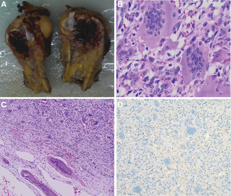
Figure 2.
Postoperative pathological and immunohistochemical staining results. 2A Mass specimen. 2B Microscopically, the mass was composed of mononuclear stromal cells and osteoclast-like giant cells. HE 400×. 2Creactive/metaplastic bone formation were seen around the lesion. HE 100×. 2DTumor cells were negative for H3.3G34W. IHC 100×.

