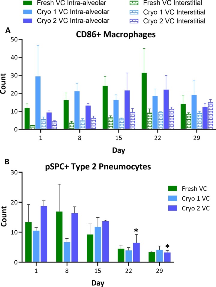
Figure 6.
Levels of intra-alveolar and interstitial CD86+ macrophages and type 2 pneumocytes. The hPCLS were stained with IHC stains for CD86 (A) or pSPC (B). The stained cells were counted in 5 high-powered fields. Changes in the levels of interstitial macrophages and type 2 pneumocytes were observed over the course of culture period. The scores are presented as AVE ± SEM, N = 3/group per time point.

