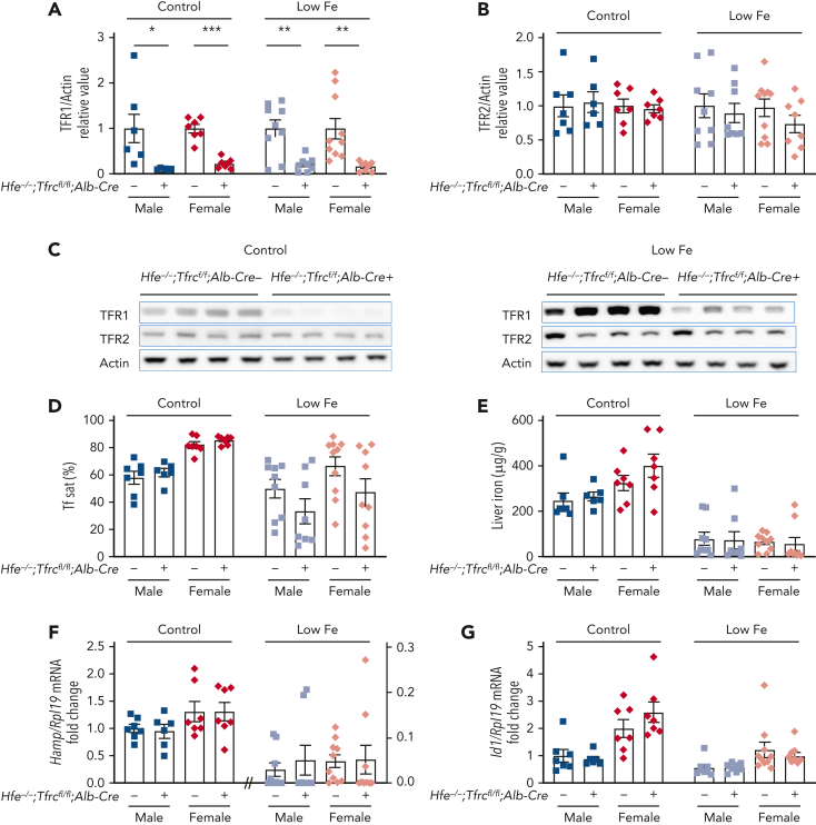
Figure 2.
Deletion of hepatocyte Tfrc in Hfe knockout mice does not alter iron phenotype compared with Hfe single knockout mice. Four to 5-week-old male (blue) and female (red) double global Hfe knockout; hepatocyte Tfrc knockout mice (Hfe−/−;Tfrcfl/fl;Alb-Cre+) and littermate single Hfe knockout mice (Hfe−/−;Tfrcfl/fl;Alb-Cre−) were either maintained on a standard diet (control, ∼380 ppm iron) or low-iron diet (low Fe, 2-6 ppm iron) for 3 weeks. Livers were analyzed for (A,C) TFR1 and (B-C) TFR2 relative to total actin protein by immunoblot and chemiluminescence quantitation. For panels A-B, the average of the Hfe−/−;Tfrcfl/fl;Alb-Cre− mice for each sex and diet was set to 1. Representative immunoblots are shown. (D) Serum transferrin saturation and (E) liver iron levels were analyzed by colorimetric assays. Livers were analyzed for (F) Hamp and (G) Id1 relative to Rpl19 mRNA by qRT-PCR. For panels F-G, the average of the Hfe−/−;Tfrcfl/fl;Alb-Cre− male mice on the standard diet was set to 1. For all graphs, individual data points are shown, and bars represent mean ± SEM. ∗P < .05, ∗∗P < .01, ∗∗∗ P < .001 relative to diet- and sex-matched Hfe−/−;Tfrcfl/fl;Alb-Cre− mice by the Student t test. qRT, quantitative reverse transcription; SEM, standard error of the mean.

