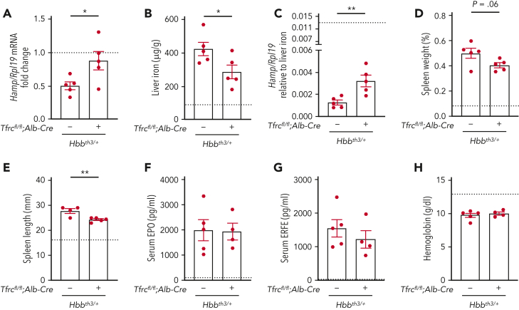
Figure 4.
Deletion of hepatocyte Tfrc ameliorates hepcidin suppression and liver iron overload in β-thalassemic mice. Six-week-old littermate female double-mutant hepatocyte Tfrc knockout thalassemia mice (Hbbth3/+;Tfrcfl/fl;Alb-Cre+), thalassemia mice (Hbbth3/+;Tfrcfl/fl;Alb-Cre−), and nonthalassemic controls (Hbb+/+;Tfrcfl/fl;Alb-Cre−, represented by dotted line) were analyzed for (A) Hamp relative to Rpl19 mRNA by qRT-PCR. The average of the nonthalassemic control mice was set to 1. (B) Liver iron levels were analyzed by colorimetric assay. (C) Hamp/Rpl19 mRNA was divided by liver iron content to normalize for the degree of iron overload. (D-E) Spleens were measured for (D) weight divided by total body weight and (E) length. (F) Serum EPO and (G) ERFE protein levels were quantified by ELISA. (H) Hemoglobin levels were analyzed by CBC. For all graphs, the average of the nonthalassemic group is shown as a dotted line. For other genotypes, individual data points are shown, and bars represent mean ± SEM. ∗P < .05, ∗∗P < .01 for Hbbth3/+;Tfrcfl/fl;Alb-Cre+ mice relative to Hbbth3/+;Tfrcfl/fl;Alb-Cre− mice by the Student t test. ELISA, enzyme-linked immunosorbent assay; qRT, quantitative reverse transcription; SEM, standard error of the mean.

