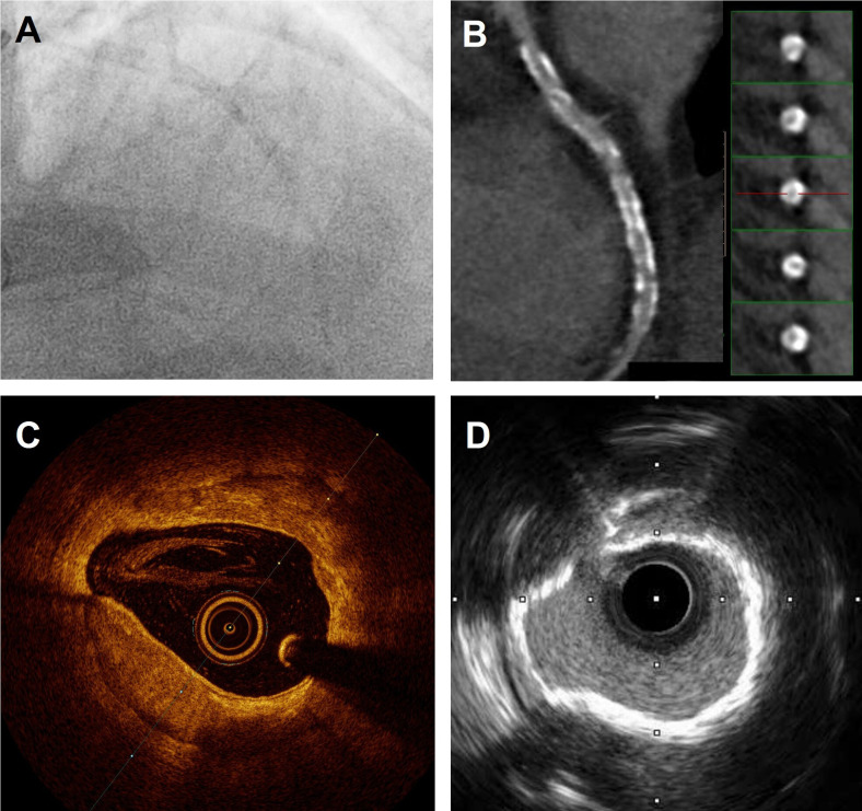
Figure 2.
Coronary calcification depicted with multimodality imaging. (A) Angiographic image showing heavily calcified left anterior descending artery (LAD). (B) CT showing widespread calcification. (C) OCT images showing deep circumferential calcium with well-defined borders. (D) IVUS imaging showing calcium ring that is highly echogenic. IVUS, intravascular ultrasound; OCT, optical coherence tomography.

