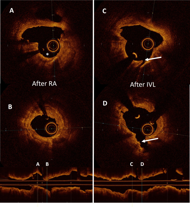
Figure 3.
Optical coherence tomography showing the results after sequential use of rotational atherectomy (RA) and intravascular lithotripsy (IVL). (A and B) Two separate cross-sectional frames of a heavily calcified lesion treated with RA. The asterisk shows the cavity formed by the pass of the burr. (C and D) cross-sectional frames after the use of a lithotripsy balloon. The arrow indicates the creation of a deep fracture in the body of the calcium deposit.

