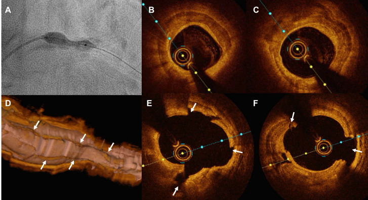
Figure 5.
Detailed depiction with optical coherence tomography (OCT) of the effects of intravascular lithotripsy throughout a heavily calcified undilatable lesion. (A) Underexpansion of a 4.0 mm high-pressure balloon in a calcified proximal left anterior descending artery. (B and C) OCT cross-sectional frames showing the presence of thick calcium sheets surrounding the vessel. (D–F) OCT three-dimensional long view reconstruction and cross-sectional frames after the use of lithotripsy showing the cracks formed in the calcium surface fracturing its body creating deep grooves.

