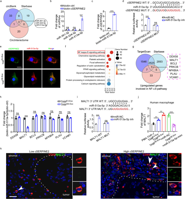
Fig. 4.
Tumor exosomal cSERPINE2 upregulated MALT1 expression in macrophages by sponging miR-513a-5p. a Identification of miRNAs that potentially bind to cSERPINE2 based on CircInteractome, circBank and Starbase databases. b RNA pull-down assay was performed in TAMOE−Exo cells using biotinylated cSERPINE2-specific probe and control probe. The enrichment of miRNAs was detected by qRT-PCR. c RNA pull-down assay was performed in TAMOE−Exo cells using biotinylated miR-513a-5p probe and control probe. The enrichment of cSERPINE2 was detected by qRT-PCR. d A schematic drawing showing the putative binding sites of miR-513a-5p in cSERPINE2 sequence, and the luciferase activity of cSERPINE2-WT or cSERPINE2-MUT in 293 T cells after co-transfection with miR-513a-5p inhibitor. e Representative FISH images of co-localization between miR-513a-5p and cSERPINE2 in TAM EV−Exo and TAM OE−Exo cells. Scale bar, 10 μm. f Pathway enrichment of the up-regulated genes in TAMOE−Exo according to KEGG analysis. g Venn diagram showing the intersections of up-regulated genes involved in the NF-B pathway and potential targets of miR-513a-5p predicted by Starbase and TargetScan. h RNA pull-down assay was performed in TAMEV−Exo and TAMOE−Exo cells using biotinylated miR-513a-5p probe and control probe. The enrichment of RNAs was detected by qRT-PCR. i A schematic drawing showing the putative binding sites of miR-513a-5p in MALT1 3’UTR sequence, and the luciferase activity of MALT1-WT or MALT1-MUT in 293 T cells after co-transfection with miR-513a-5p inhibitor. j The relative expression of MALT1 in PMA-THP-1 and TAMs as indicated treatments. k Immunofluorescence showing MALT1 expression (Green) on CD68+ macrophages (Red) in breast cancer tissues with low or high cSERPINE2 expression. Scale bar, 20 μm. Data are presented as the means ± SD of three independent experiments. *P < 0.05, **P < 0.01, ***P < 0.001

