Fig. 2.
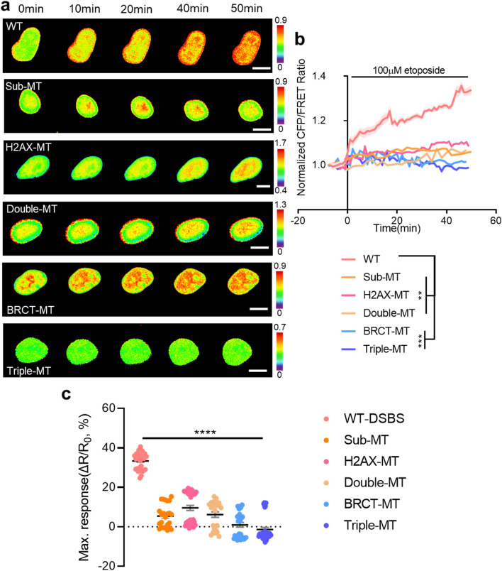
Dynamic visualization of the DSBS in response to etoposide-induced DSBs. a Time-lapse CFP/FRET ratio images of the WT-DSBS, Sub-MT, H2AX-MT, Double-MT, BRCT-MT, and Triple-MT in HEK293T cells treated with 100 μM etoposide. The color scale indicates high (red) and low (blue) levels of DSBs (scale bar = 10 μm). b Time courses of the normalized CFP/FRET ratio of the WT-DSBS (n = 5) and mutants (n = 4–5) in HEK293T cells treated with 100 μM etoposide. c The maximum responses of the biosensors in HEK293T cells treated with 100 µM etoposide (n = 24–45). The ratio changes (∆R, Rmax-R0) were divided by the basal ratio (R0), where Rmax is the maximum ratio after etoposide treatment. All error bars represent SEM (**P < 0.01, ***P < 0.001, ****P < 0.0001)

