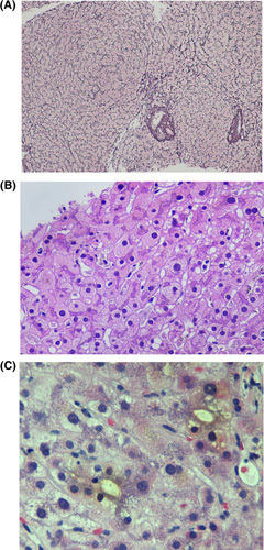
FIGURE 2.

Examples of histological injury attributed to DILI. (A) Nodular regenerative hyperplasia can be seen with azathioprine and oxaliplatin. Reticulin stain highlights a nodular architecture with nodules made up of hyperplastic hepatocytes characterized by two‐cell‐thick plates that are bordered by atrophic hepatocyte plates. Note that the portal tract (arrow) is in the center of the nodule, termed reverse lobulation (original magnification ×10, reticulin stain). (B) Hepatocytes with ground‐glass like cytoplasm are characterized by smooth homogeneous light pink color as opposed to the typical grainy eosinophilic cytoplasm of normal hepatocytes. These hepatocytes are typically found in zone 3. The development of these is often due to polypharmacy (original magnification ×10, hematoxylin and eosin stain). (C) Photomicrograph showing dilated canaliculi containing bile but no inflammatory infiltrates are present and very rare hepatocytes are noted to be undergoing feathery degeneration. This pattern of injury is reported with drugs such as trimethoprim‐sulfamethoxazole (original magnification ×40, hematoxylin and eosin).
