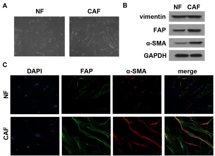
FIG 1.
CAFs were successfully isolated. (A) The morphology of the isolated CAFs and NFs was observed under an inverted microscope. (B) Expression of signature markers α-SMA, FAP and vimentin were determined by Western blot analysis. (C) Immunofluorescence staining was applied to assess the abundance of α-SMA and FAP in CAFs and NFs.

