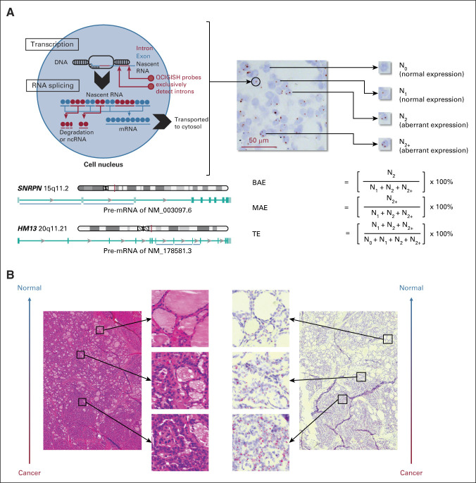
FIG 2.
The principle of QCIGISH—visualization, quantification, and pathologic confirmation of the allelic expression status of imprinted genes. (A) Conceptual framework of QCIGISH. Shown is a QCIGISH-stained tissue section of a case of PTC. Blue components in the image are cell nuclei stained using hematoxylin. The red vertical lines on the chromosome map indicate the gene loci. The blue horizontal lines under the intron/exon map of pre-mRNAs indicate the targeted introns of the in situ hybridization probes. (B) Pathologic confirmation of the QCIGISH results using simultaneous hematoxylin and eosin staining examination of a case of PTC. The low-magnification image was captured at a particular tumor region showing both malignant (lower subregion) and benign (upper subregion) morphologic characteristics. BAE, biallelic expression; MAE, multiallelic expression; ncRNA, noncoding RNA; PTC, papillary thyroid carcinoma; QCIGISH, quantitative chromogenic imprinted gene in situ hybridization; TE, total expression.

