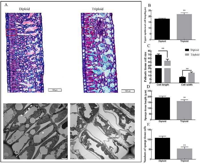
Fig 3. Transverse section of tea diploid and triploid leaf mesophyll.
A: Diploid and triploid mesophyll cross sections under 100X magnification. B-E: Cross sectional length of upper epidermal cell, size of the palisade tissue cell, number of sponge tissue cells and sponge tissue length. Error bars indicate SD (n = 3); ** indicates significant difference at the 0.01 level between the diploid and triploid.

