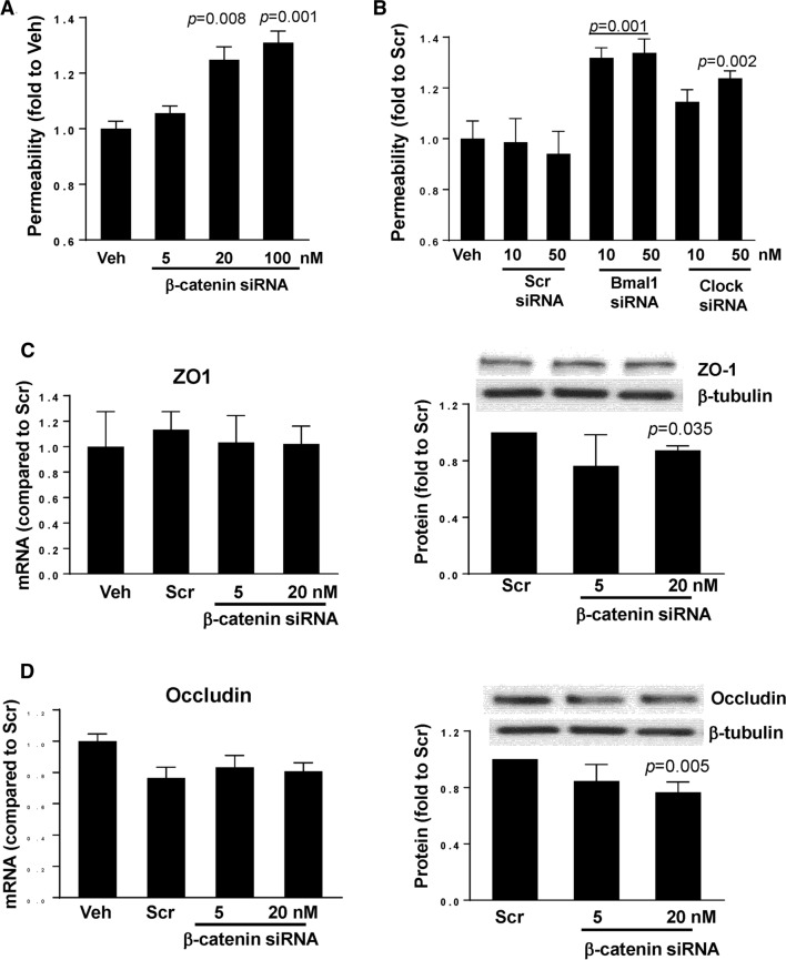
Fig. 6.
Silencing of circadian clock molecules increases paracellular permeability across epithelial monolayers. SW480 cells grown on the apical side of wells (0.4-μm pore size, 12-mm diameter) of Transwell inserts were transfected with siRNA of β-catenin (A), Bmal1, or Clock (B) at the indicated concentrations. Control cultures were either treated with vehicle (Veh) or transfected with nonspecific, scrambled (Scr) siRNA. Epithelial permeability was determined by measuring paracellular passage of FITC-dextran 20 kDa from the apical side to the basolateral side across SW480 cell layers. A p indicates the level of statistical significance after β-catenin silencing as compared to the Veh group. B p indicates the level of statistical significance after Bmal1 or Clock silencing as compared to the Veh or Scr group. Values (means ± SD) are expressed as fold change compared with Veh or Scr. Values are mean ± SEM; n = 5–6. C, D Silencing of β-catenin alters expression of tight junction proteins without affecting mRNA levels. SW480 cells grown on the 12 well cell culture plates were transfected with β-catenin-specific siRNA at the indicated concentration or with nonspecific, scrambled siRNA. mRNA (left panels) or protein (right panels) expression of ZO-1 (C) and occludin (D) were assayed and quantified. Blots illustrate representative data. Band intensity was assessed by densitometric analysis using Image J program. p indicates the level of statistical significance after β-catenin silencing as compared to the Scr group. Values (means ± SD) are expressed as fold change compared with Scr; n = 3–6

