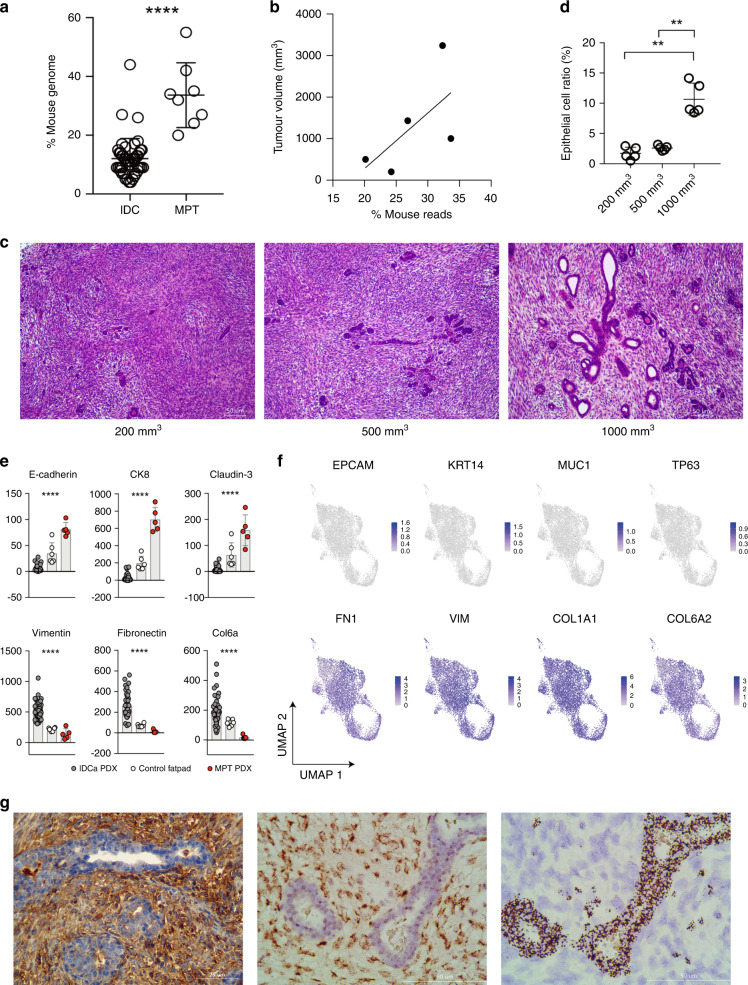
Fig. 4. Role of epithelial cells and stromal cells in malignant phyllodes tumours (MPTs).
a Proportion of RNA-seq reads originating from murine cells in PDX derived from invasive ductal carcinoma (IDC) and MPT (Mann–Whitney test). b Correlation between MPT tumour volume and the proportion of RNA-seq reads originating from murine cells in the MPT PDX. c Haematoxylin and eosin staining results of MPT PDX tumour resected at volumes 200 mm3 (left), 500 mm3 (middle), and 1000 mm3 (right). d Proportion of epithelial cells according to tumour volume (Mann–Whitney test). e Expression levels of epithelial cell markers (top line) and mesenchymal stromal cell markers (bottom line) based on mouse transcriptome in normal mouse fat pads and PDX derived from MPT and IDC (Kruskal–Wallis test). f UMAP embeddings of epithelial markers expressing grafted human tumour cells (top) and fibroblast markers expressing grafted human tumour cells (bottom) (n = 8105). g Identification of human mesenchymal cells and mouse epithelial cells by anti-human HLA class 1 staining (left), anti-FITC staining after human-specific centromeric FISH (middle), and mouse-specific centromeric FISH (right). **p < 0.01, ****p < 0.0001.

