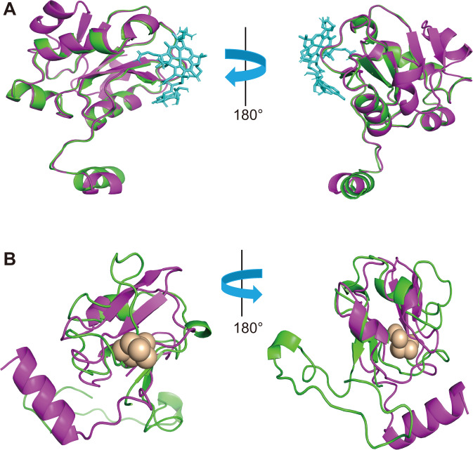
Fig. 3. Structure comparison between HgcA and HgcB from Lokiarchaeota and Desulfovibrio desulfuricans ND132.
A Superposition of Lokiarcheaota-HgcA (green) and Desulfovibrio desulfuricans ND132-HgcA (purple) functional domain homology models. The functional domain is bound to cobalamin shown cyan-blue stick; B Superposition of Lokiarchaeota-HgcB (green) and Desulfovibrio desulfuricans ND132-HgcB (purple) homology models. Model of full-length Lokiarchaeota-HgcB in complex with two [4Fe-4S] clusters; Interactions between the protein and iron are shown by brown ball.

