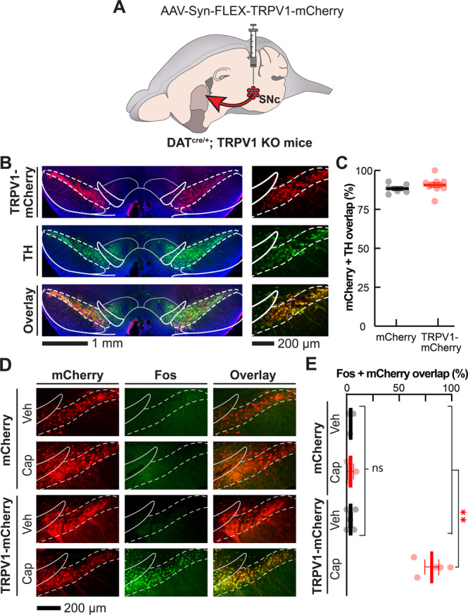
Fig. 1. Histological validation of the selective expression of functional TRPV1 in SNc dopamine neurons.

A We virally expressed Cre-dependent mCherry (control) or TRPV1-mCherry (experimental) in DS-projecting SNc dopamine neurons of DATcre/+; TRPV1 KO mice. B Coronal brain sections containing SNc and VTA from a representative experimental (TRPV1) mouse shown at 10× (left) and 16× resolution (right; blue: DAPI nuclear stain; red: α-RFP; green: α-TH). C Mean ± s.e.m. percentage of mCherry-expressing neurons that are TH positive in control and TRPV1 mice (N = 6 and 10 respectively; average of 3 brain slices per mouse). D Coronal brain sections containing SNc from representative control (top) and TRPV1 (bottom) mice immunostained for Fos (green) and RFP (red) 75 min after systemic vehicle or capsaicin treatment (10 mg·kg−1). E Mean ± s.e.m. percentage of mCherry-expressing neurons that are Fos positive as a function of treatment and experimental group (N = 3–5 mice per group; average of 3 brain slices per mouse). In all images, white dashed lines indicate boundaries of SNc and solid lines indicate boundaries of adjacent brain structures. **P < 0.01 comparing treatments and groups; Kruskal-Wallis unpaired one-way ANOVA in (E). Details for these and all other statistical comparisons are presented in the Supplementary Table.
