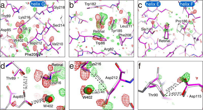
Fig. 3. Propagation of structural changes from retinal to the protein matrix.
a A cross-section of retinal at the SB linkage. The F(K + bR) − F(bR) difference map is shown as green and red meshes at contour levels of ±3σ. The K structures are represented as colored sticks. The ground state structure is overlaid as gray sticks. b A cross-section of retinal at the middle of the polyene backbone. c A cross-section of retinal at the β-ionone ring. d The map around Asp85. e The map around Asp212. f The map around Asp115.

