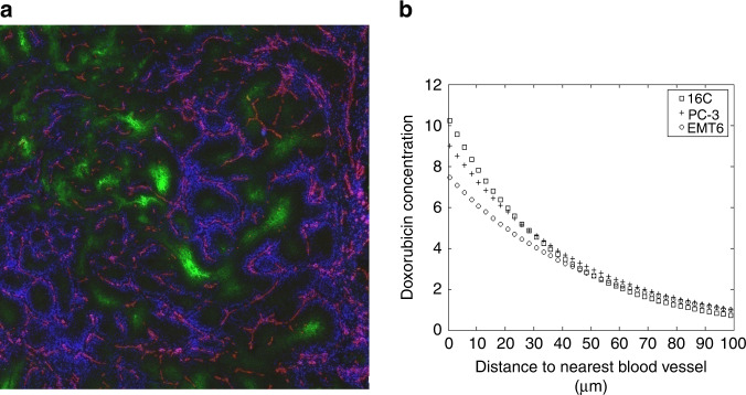
Fig. 2. Distribution of doxorubicin in solid tumours.
a Distribution of fluorescent doxorubicin (false-colour blue) around blood vessels (recognised by CD31 on endothelial cells: false-colour red) in a mouse mammary tumour. Hypoxic areas are recognised by binding to EF5 (false-colour green). b Doxorubicin fluorescence as a function of distance from the nearest blood vessel in sections from three mouse mammary tumours. Modified from Primeau et al. [16].

