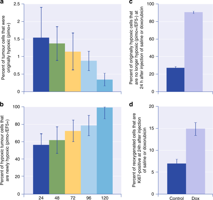
Fig. 3. Time-dependent changes in hypoxic cells, and in their proliferation, in doxorubicin-treated and control mice bearing MCF-7 xenografts.
Mice were injected with 2 hypoxic cell markers (pimonidazole and EF5) with a variable interval between. a Percent of tumour cells that were originally hypoxic (pimo + ) as a function of time. b Percent of hypoxic tumour cells that are newly hypoxic (pimo−/EF5 + ) as a function of time. There is dynamic flux of cells through the hypoxic compartment. c Percent of originally hypoxic cells that are no longer hypoxic (pimo + /EF5−) at 24 h after injection of saline or doxorubicin. d Percent of reoxygenated cells that are Ki67 positive at 24 h after injection of saline or doxorubicin. Reoxygenation and repopulation of hypoxic cells occur after treatment with doxorubicin. Similar results were obtained after treatment of prostate cancer PC3 xenografts with docetaxel. Adapted from Saggar and Tannock [18].

