Figure 1:

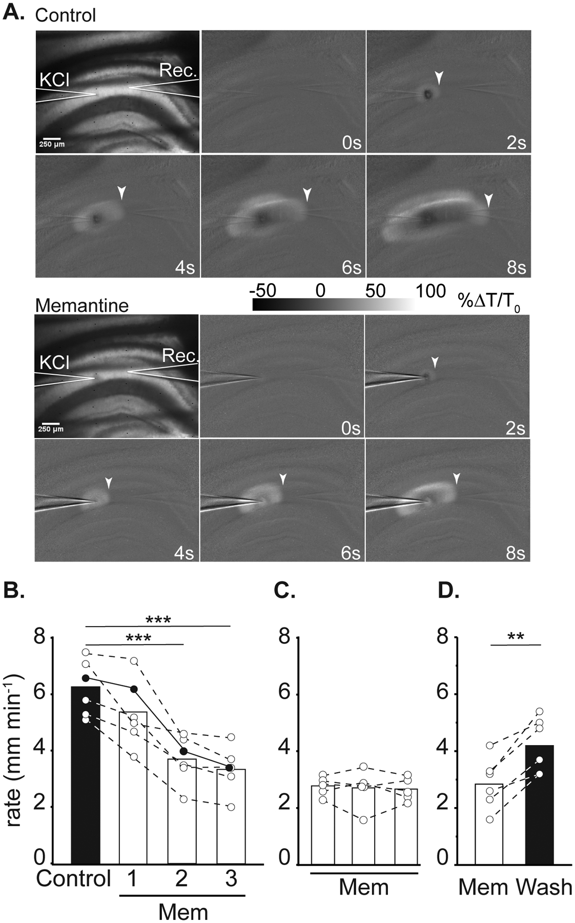
Effect of memantine on SD propagation rate. A: Representative images demonstrating electrode placements (KCl, Rec.) and changes in transmitted light during SD propagation in control (top panel) and in memantine (below, 3rd SD in mem) within the same slice. Arrowheads indicate the wavefront of SD, scale bar = 250μm. B: Summary data showing SD propagation rates during successive SD stimulations from three separate sets of experiments. Left: effect of acute memantine wash in (n = 6) on SD rate, filled symbols are values measured from the preparation shown in A. *** P<0.001 compared to the second control SD. Middle: slower propagation rates during the first SD following extended memantine exposure (≥ 3h, n = 5). Right: faster SD propagation could be observed following extensive memantine wash out (n = 6). **P<0.01 third SD in memantine compared to wash out.
