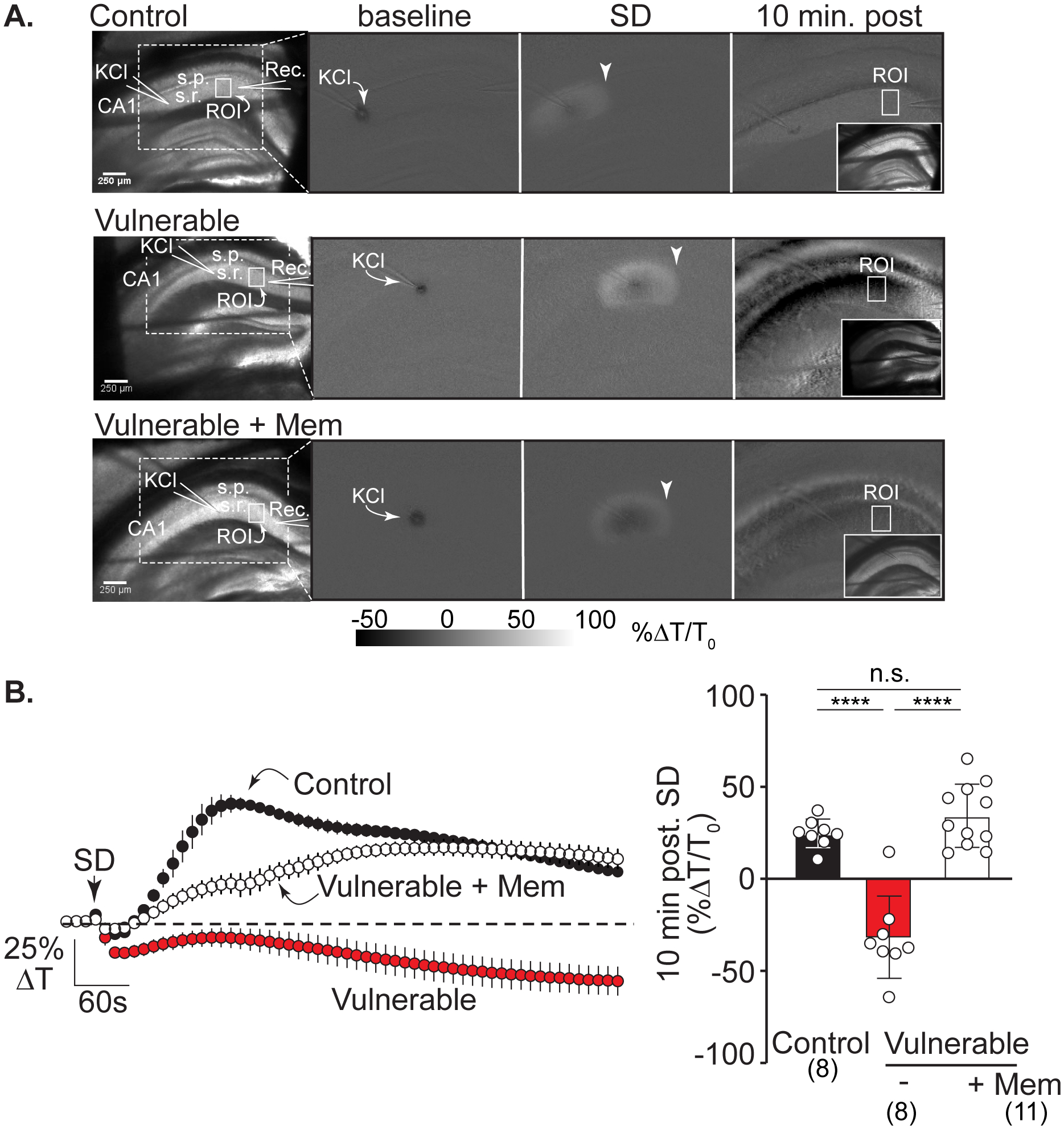
Figure 4:

Memantine protects against IOS decreases after SD in vulnerable brain slices. A: Left hand panel shows the arrangement of KCl and recording electrodes in transmitted light images from representative slices in the three experimental conditions: control, vulnerable, and vulnerable + memantine (>2h). Solid white boxes show the regions of interest (ROIs) positioned in CA1 stratum radiatum and used for analyses; s.r.: stratum radiatum, s.p.: stratum pyramidale, scale bar = 250μm. The white dotted box outlines the imaging area shown in subsequent panels demonstrating changes in intrinsic optical signals (IOS, light transmittance) at baseline (during KCl microinjection), during SD propagation (arrowheads), and at the 10-minute time point after SD. Right hand inset panels show transmitted light images of each slice 10 minutes post SD. In control recording conditions (top row) SD led to a persistent IOS increase in stratum radiatum regions, consistent with full recovery in these nominally healthy conditions. In contrast, in the vulnerable conditions (middle row) there was persistent decrease IOS after SD, characteristic of lack of recovery. Pre-exposure to memantine (bottom row) prevented the decrease in IOS signal, consistent with improved recovery of SD in these metabolically compromised conditions. B: Average IOS traces (left; from n = 27 preparations) extracted from regions in CA1 (ROIs in stratum radiatum shown in A) during experiments shown in A. Summary data (right) confirm substantial decreases in CA1 light transmittance 10 minutes after passage of the SD wavefront in vulnerable slices (red symbols, n = 8) compared to control conditions (black symbols n = 8) with prevention by memantine pretreatment (>2h, white symbols, n = 11). ****P<0.0001
